Neurology - Blog Posts
Just made an account on Medium so I could read this. Made it about 1/3 through and I'm absolutely hooked and excited to finish it!! But first, time for a nap.
For the longest time, I have had trouble understanding FND - specifically whether it's just a shitty diagnosis made up by the medical system to gaslight people or if it's a genuine medical condition. This essay is rly helpful and has already broadened my understanding. It's incredibly well written, particularly bc of the extremely thorough research it's a result of

I am one of the most medically examined people in North America. For over a decade, no one could explain why I lost my ability to walk, speak, and use my hands. Why the lightning-like headaches? Why the ringing in my ears? Test after test came back negative. Doctors thought I might have a genetic abnormality no one's ever seen before, or a condition so rare that it had previously escaped medical classification. Then I got accepted to the top undiagnosed disease research program in the world, and they told me the only diagnosis I was unprepared to hear: it was Functional Neurological Disorder (FND), a much-misunderstood condition which was once known as Conversion Disorder, and before that, as Hysteria. And that was only the beginning of things getting weird. The essay above is the product of three years of research into the history, neuroscience, and politics of FND. It touches on the many medical failures that define the history of the disorder, the pervasive sexism and lazy mind-body dualism that prevented scholars from seeing it clearly, and why - finally - a better understanding may be at hand, with revolutionary implications for how we understand human consciousness and the experience of having a body.
FND fucked my life up. This is my reply. Thanks for reading.
✨molecular naturecore.✨

Scan of 1 cubic millimeter of the human brain
Full scan of 1 cubic millimeter of brain tissue that took 1.4 petabytes of data, equivalent to 14,000 4K movies.
Slime Molds and Intelligence

Okay, despite going into a biology related field, I only just learned about slime molds, and hang on, because it gets WILD.
This guy in the picture is called Physarum polycephalum, one of the more commonly studied types of slime mold. It was originally thought to be a fungus, though we now know it to actually be a type of protist (a sort of catch-all group for any eukaryotic organism that isn't a plant, animal, or a fungus). As protists go, it's pretty smart. It is very good at finding the most efficient way to get to a food source, or multiple food sources. In fact, placing a slime mold on a map with food sources at all of the major cities can give a pretty good idea of an efficient transportation system. Here is a slime mold growing over a map of Tokyo compared to the actual Tokyo railway system:

Pretty good, right? Though they don't have eyes, ears, or noses, the slime molds are able to sense objects at a distance kind of like a spider using tiny differences in tension and vibrations to sense a fly caught in its web. Instead of a spiderweb, though, this organism relies on proteins called TRP channels. The slime mold can then make decisions about where it wants to grow. In one experiment, a slime mold was put in a petri dish with one glass disk on one side and 3 glass disks on the other side. Even though the disks weren't a food source, the slime mold chose to grow towards and investigate the side with 3 disks over 70% of the time.

Even more impressive is that these organisms have some sense of time. If you blow cold air on them every hour on the hour, they'll start to shrink away in anticipation when before the air hits after only 3 hours.
Now, I hear you say, this is cool and all, but like, I can do all those things too. The slime mold isn't special...
To which I would like to point out that you have a significant advantage over the slime mold, seeing as you have a brain.
Yeah, these protists can accomplish all of the things I just talked about, and they just... don't have any sort of neural architecture whatsoever? They don't even have brain cells, let alone the structures that should allow them to process sensory information and make decisions because of it. Nothing that should give them a sense of time. Scientists literally have no idea how this thing is able to "think'. But however it does, it is sure to be a form of cognition that is completely and utterly different from anything that we're familiar with.

Enjoy this First-Year-Anniversary compilation of all of my works in one title: A Cosmic Legacy: From Earth to the Stars This title includes the following works wrapped up into one story: Further Than Before: Pathway to the Stars, Part 1 Further Than Before: Pathway to the Stars, Part 2 Pathway to the Stars: Part 1, Vesha Celeste Pathway to the Stars: Part 2, Eliza Williams Pathway to the Stars: Part 3, James Cooper Pathway to the Stars: Part 4, Universal Party Pathway to the Stars: Part 5, Amber Blythe Pathway to the Stars: Part 6, Erin Carter "Our beautiful mother world ached for a reprieve from the injustices of many, courtesy of cultures and governance systems, that forgot how to love, how to be kind, how to include others, and how to think beyond the scope of greed and power, but within the visions of shared joy and well-being." Together with the organization Eliza Williams founded, called Pathway, she and her growing team will take us on a fantastical and Utopian journey to get us out and into the farthest reaches of space. There are dilemmas such as the physiological effects of space on each of us, as well as the need for longevity and a desire to still be able to visit loved ones following long journeys. Eliza and her team develop capabilities, so we can overcome the challenges ahead and are determined to stabilize a rocky economy, wipe away suffering, violence, disease, cartels, terrorism, and trafficking in persons. They work together to tame seismic activity, weather, and fires. She and her friends tackle ways to prevent extinction and provide solutions to quality of life concerns. They even consider the longevity of our Sun and our Earth's capacity to preserve life. Eliza tackles each of these issues to get us out, and into the stars, so we can begin our biggest quest--to help our Universe breathe ever so lightly. #amazing #science #fiction #novels #best #new #books #scifi #online #read #longevity #CRISPR #physiology #neurology #physics #theoretical #philosphical #politcal #educational #STEM #AmazonAuthor #BarnesAndNobleAuthor #wellbeing #quality #biotech #nanotech #SpaceOpera #astronomy #selfpublished https://www.instagram.com/p/B2GkDbYBs0y/?igshid=ufavr7j6lsy1

Curl up, read a new series of books, and be edified! Out now, Part 1, Vesha Celeste, and Part 2, Eliza Williams, have been paired together, and are available for those interested in the types and directions of science and the speculation that lead to well-being and quality of life. Please feel free to follow, message, share ideas, and be a part of a positive future where, if we choose, we can prepare properly to navigate the stars. This is just the beginning of this series and prequel, “Pathway to the Stars,” to an even more giant series, “Further than Before!” Please enjoy. https://www.amazon.com/author/matthewopdyke #sciencefiction #scifi #sciencefictionfantasy #scififantasy #politicalscifi #physiology #neurology #physics #Apolitical #strongfemalelead #biotechnology #neuroscience #theoreticalphysics #problemresolution #productivepursuits #spaceopera #cerebraldrama #sophisticateddialogue #solarsystem #spacemining #physiologicaloptimization, #neurologicaloptimization #transhumanism #universalethics #wellbeing #genetherapy #CRISPR #politicalsciencefiction #matthewopdyke https://www.instagram.com/p/BpUMW6ZgB1I/?utm_source=ig_tumblr_share&igshid=wy078mgrbgmm
Covid Findings, a Lay Person’s Account.
Hello,
I have popped back to share my own personal experience, and to help heal myself after watching a snippet of a recent BBC Question Time television programme, which I felt only served to try to shame people who were making their own free choices as a human being.
Just a small snippet, where there first guest did not appear to be able to articulate well what he was saying, and had made an unfortunate mistake in a fact (perhaps a victim of actual disinformation), and a second guest who got all of her valid points across, only to be shamed for it on national television in front of a panel of professionals? I thought that the days of barbarianism were over in the UK at least, yet here they are still today only in a different format on television.
I felt like I had just watched a small snippet of abuse, unfortunately this is spread over an entire length of a program involving many more guests than the ones that I have mentioned. It was painful to watch, and I felt that it was aiming to get the monetary audiences in, not just those present to discuss.
I have never spoken about my own experiences with Covid-19, I wanted to ensure that I was not influencing anyone else’s freedoms, but since the BBC are okay to try and shape peoples opinions that I at least should be permitted to write what I am about to write.
At this point, which is Friday 4th of February, 2022, and after hearing many different peoples experiences as well as re-experiencing the same, slight differing problem over the years myself, I can feel safe in the knowledge that I first came across Covid-19 in whatever form that it was in, in November / December 2015.
I then picked up the same thing in February 2016, and lasted a bit longer until I got it again in February 2020, March 2020, October 2020, June 2021, and January 2022 - the recent episodes in January lasted me two days at most, and was not as rough terrain like previous experiences.
Throughout that time I had no help or understanding as to what was going on, until 2021 when I began to wonder if what I had been troubled with all this time was in fact Covid-19, so I started listening to others experiences and keeping up with what was happening for people globally in the news.
There were a few things that stood out to me along that journey;
* the affect that Covid-19 was having on peoples gut
* the re-circulation of old or dormant virus and associated symptoms
* the ability of Covid-19 to cross the brain barrier
Having plenty of time on my hands during lock down, I certainly kept my eye on the world news for updates and noticed more and more that symptoms of things like long Covid fatigue, matched with those of the known Epstein Barr virus, which is common and can lay harmlessly dormant in the guts of anyone.
I also noticed that fungus was a big player in Covid-19 mortality, as fungus is one of the causes of pneumonia. Various fungus can also be the cause of many rash like symptoms that people experience.
In looking at my own experience at least, fungus and bacteria are not just big players in Covid-19 symptoms, but the main ones, leading me to personally (as a lay person) come to understand that Covid-19 is likely a liberator of whatever lays dormant, and perhaps not so dormant, in the guts of the host that it comes to exploit. This includes the transportation and crossing of some of those things over the brain barrier, which explains why so many accounts of Covid-19 experiences include those of neurological symptoms.
Like any public transport service, Covid-19 is nothing without it’s passengers. Taking care of our general health and reducing susceptibility to the overgrowth, overexposure, and resistance to overcoming various fungus and bacteria that are naturally occurring in ourselves and in the environment, may well help in promoting the permanent closure of Covid-19′s business.
The experiences of people matter, as does the free choice for people to have a vaccine, or to not have a vaccine.
Stay well :)

Why do so many neurologists suck bro. I'm so glad I'm getting a new one

After a short break, keep working again!
I won't be taking classes because I'm in an internship this semester, but I'm still working in two labs and the obligation to do so is increasing :')
Be prepared to see posts full of articles, even though I'm still not ready to read..
For music: Hakim Bey
{Ladin}

Did you know that flies can see their surroundings at 330 degrees thanks to the lenses in their eyes? I am more surprised when I see the fly hitting everywhere in the house.
The day before the Behavioral Neurobiology exam..
{Ladin}
mossy fiber butons.
I love them. absolutely gorgeous little dudes. I mean look at em.

In the hippocampus, they typically play a key role in plasticity, processing, and encoding of information. They are also located in the cerebellum as well. I will probably post more about them as there is a ton to cover!
Brand new video about some basic stuff.
Brain things, science stuff, discussing more about hiking & almost dying (not really I had it under control but...you know it could have gone another way)
New production by Salty Wave
Salty Wave has been busy producing a series of Video Explainers for the Institute of Molecular Bioscience at the University of Queensland, Australia.
Take a look at The Science of Acute Pain to understand how pain is felt and experienced by many people.

The late effects of stress: New insights into how the brain responds to trauma
Mrs. M would never forget that day. She was walking along a busy road next to the vegetable market when two goons zipped past on a bike. One man’s hand shot out and grabbed the chain around her neck. The next instant, she had stumbled to her knees, and was dragged along in the wake of the bike. Thankfully, the chain snapped, and she got away with a mildly bruised neck. Though dazed by the incident, Mrs. M was fine until a week after the incident.
Then, the nightmares began.
She would struggle and yell and fight in her sleep every night with phantom chain snatchers. Every bout left her charged with anger and often left her depressed. The episodes continued for several months until they finally stopped. How could a single stressful event have such extended consequences?
A new study by Indian scientists has gained insights into how a single instance of severe stress can lead to delayed and long-term psychological trauma. The work pinpoints key molecular and physiological processes that could be driving changes in brain architecture.
The team, led by Sumantra Chattarji from the National Centre for Biological Sciences (NCBS) and the Institute for Stem Cell Biology and Regenerative Medicine (inStem), Bangalore, have shown that a single stressful incident can lead to increased electrical activity in a brain region known as the amygdala. This activity sets in late, occurring ten days after a single stressful episode, and is dependent on a molecule known as the N-Methyl-D-Aspartate Receptor (NMDA-R), an ion channel protein on nerve cells known to be crucial for memory functions.
The amygdala is a small, almond-shaped groups of nerve cells that is located deep within the temporal lobe of the brain. This region of the brain is known to play key roles in emotional reactions, memory and making decisions. Changes in the amygdala are linked to the development of Post-Traumatic Stress Disorder (PTSD), a mental condition that develops in a delayed fashion after a harrowing experience.
Previously, Chattarji’s group had shown that a single instance of acute stress had no immediate effects on the amygdala of rats. But ten days later, these animals began to show increased anxiety, and delayed changes in the architecture of their brains, especially the amygdala.
“We showed that our study system is applicable to PTSD. This delayed effect after a single episode of stress was reminiscent of what happens in PTSD patients,” says Chattarji. “We know that the amygdala is hyperactive in PTSD patients. But no one knows as of now, what is going on in there,” he adds.
Investigations revealed major changes in the microscopic structure of the nerve cells in the amygdala. Stress seems to have caused the formation of new nerve connections called synapses in this region of the brain. However, until now, the physiological effects of these new connections were unknown.
In their recent study, Chattarji’s team has established that the new nerve connections in the amygdala lead to heightened electrical activity in this region of the brain.
“Most studies on stress are done on a chronic stress paradigm with repeated stress, or with a single stress episode where changes are looked at immediately afterwards – like a day after the stress,” says Farhana Yasmin, one of the Chattarji’s students. “So, our work is unique in that we show a reaction to a single instance of stress, but at a delayed time point,” she adds.
Furthermore, a well-known protein involved in memory and learning, called NMDA-R has been recognised as one of the agents that bring about these changes. Blocking the NMDA-R during the stressful period not only stopped the formation of new synapses, it also blocked the increase in electrical activity at these synapses.
“So we have for the first time, a molecular mechanism that shows what is required for the culmination of events ten days after a single stress,” says Chattarji. “In this study, we have blocked the NMDA Receptor during stress. But we would like to know if blocking the molecule after stress can also block the delayed effects of the stress. And if so, how long after the stress can we block the receptor to define a window for therapy,” he adds.
Chattarji’s group first began their investigations into how stress affects the amygdala and other regions of the brain around ten years ago. The work has required the team to employ an array of highly specialised and diverse procedures that range from observing behaviour to recording electrical signals from single brain cells and using an assortment of microscopy techniques. “To do this, we have needed to use a variety of techniques, for which we required collaborations with people who have expertise in such techniques,” says Chattarji. “And the glue for such collaborations especially in terms of training is vital. We are very grateful to the Wadhwani Foundation that supports our collaborative efforts and to the DBT and DAE for funding this work,” he adds.
Aphasia: The disorder that makes you lose your words

It’s hard to imagine being unable to turn thoughts into words. But, if the delicate web of language networks in your brain became disrupted by stroke, illness or trauma, you could find yourself truly at a loss for words. This disorder, called “aphasia,” can impair all aspects of communication. Approximately 1 million people in the U.S. alone suffer from aphasia, with an estimated 80,000 new cases per year. About one-third of stroke survivors suffer from aphasia, making it more prevalent than Parkinson’s disease or multiple sclerosis, yet less widely known.

There are several types of aphasia, grouped into two categories: fluent (or “receptive”) aphasia and non-fluent (or “expressive”) aphasia.

People with fluent aphasia may have normal vocal inflection, but use words that lack meaning. They have difficulty comprehending the speech of others and are frequently unable to recognize their own speech errors.

People with non-fluent aphasia, on the other hand, may have good comprehension, but will experience long hesitations between words and make grammatical errors. We all have that “tip-of-the-tongue” feeling from time to time when we can’t think of a word. But having aphasia can make it hard to name simple everyday objects. Even reading and writing can be difficult and frustrating.

It’s important to remember that aphasia does not signify a loss in intelligence. People who have aphasia know what they want to say, but can’t always get their words to come out correctly. They may unintentionally use substitutions, called “paraphasias” – switching related words, like saying dog for cat, or words that sound similar, such as house for horse. Sometimes their words may even be unrecognizable.

So, how does this language-loss happen? The human brain has two hemispheres. In most people, the left hemisphere governs language. We know this because in 1861, the physician Paul Broca studied a patient who lost the ability to use all but a single word: “tan.” During a postmortem study of that patient’s brain, Broca discovered a large lesion in the left hemisphere, now known as “Broca’s area.” Scientists today believe that Broca’s area is responsible in part for naming objects and coordinating the muscles involved in speech. Behind Broca’s area is Wernicke’s area, near the auditory cortex. That’s where the brain attaches meaning to speech sounds. Damage to Wernicke’s area impairs the brain’s ability to comprehend language. Aphasia is caused by injury to one or both of these specialized language areas.
Fortunately, there are other areas of the brain which support these language centers and can assist with communication. Even brain areas that control movement are connected to language. Our other hemisphere contributes to language too, enhancing the rhythm and intonation of our speech. These non-language areas sometimes assist people with aphasia when communication is difficult.

However, when aphasia is acquired from a stroke or brain trauma, language improvement may be achieved through speech therapy. Our brain’s ability to repair itself, known as “brain plasticity,” permits areas surrounding a brain lesion to take over some functions during the recovery process. Scientists have been conducting experiments using new forms of technology, which they believe may encourage brain plasticity in people with aphasia.
Meanwhile, many people with aphasia remain isolated, afraid that others won’t understand them or give them extra time to speak. By offering them the time and flexibility to communicate in whatever way they can, you can help open the door to language again, moving beyond the limitations of aphasia.
Neuro chip records brain cell activity at higher resolution
Brain functions are controlled by millions of brain cells. However, in order to understand how the brain controls functions, such as simple reflexes or learning and memory, we must be able to record the activity of large networks and groups of neurons. Conventional methods have allowed scientists to record the activity of neurons for minutes, but a new technology, developed by University of Calgary researchers, known as a bionic hybrid neuro chip, is able to record activity in animal brain cells for weeks at a much higher resolution. The technological advancement was published in the journal Scientific Reports.

“These chips are 15 times more sensitive than conventional neuro chips,” says Naweed Syed, PhD, scientific director of the University of Calgary, Cumming School of Medicine’s Alberta Children’s Hospital Research Institute, member of the Hotchkiss Brain Institute and senior author on the study. “This allows brain cell signals to be amplified more easily and to see real time recordings of brain cell activity at a resolution that has never been achieved before.”
The development of this technology will allow researchers to investigate and understand in greater depth, in animal models, the origins of neurological diseases and conditions such as epilepsy, as well as other cognitive functions such as learning and memory.
“Recording this activity over a long period of time allows you to see changes that occur over time, in the activity itself,” says Pierre Wijdenes, a PhD student in the Biomedical Engineering Graduate Program and the study’s first author. “This helps to understand why certain neurons form connections with each other and why others won’t.”
The cross-faculty team created the chip to mimic the natural biological contact between brain cells, essentially tricking the brain cells into believing that they are connecting with other brain cells. As a result, the cells immediately connect with the chip, thereby allowing researchers to view and record the two-way communication that would go on between two normal functioning brain cells.
“We simulated what Mother Nature does in nature and provided brain cells with an environment where they feel as if they are at home,” says Syed. “This has allowed us to increase the sensitivity of our readings and help neurons build a long-term relationship with our electronic chip.”
While the chip is currently used to analyze animal brain cells, this increased resolution and the ability to make long-term recordings is bringing the technology one step closer to being effective in the recording of human brain cell activity.
“Human brain cell signals are smaller and therefore require more sensitive electronic tools to be designed to pick up the signals,” says Colin Dalton, adjunct professor in the Department of Electrical and Computer Engineering at the Schulich School of Engineering and a co-author on this study. Dalton is also the facility manager of the University of Calgary’s Advanced Micro/nanosystems Integration Facility (AMIF), where the chips were designed and fabricated.
Researchers hope the technology will one day be used as a tool to bring personalized therapeutic options to patients facing neurological disease.
People who have any physical issues, I've created a discord server for you all! Hope that you'll have fun!
https://discord.com/invite/MgU9nvnK


Memory Competition
Most of the brain contains cells that no longer divide and renew. However, the dentate gyrus, nestled within the memory-forming centre of the brain (the hippocampus) is one of the few sites where new cells continue to form throughout life. As a person ages, there is an ever-increasing struggle for these new dentate gyrus neurons (coloured pink) to integrate with existing older neurons (green) because the latter already has well-established connections. This may be why learning and memorisation becomes more difficult as a person gets older. Scientists have now found that by temporarily reducing the number of dendritic spines – branches of neurons that form connections with other neurons – in the mature cells, the new cells have a better chance of functionally integrating. Indeed, in live mice, briefly eliminating dendritic spines boosted the number of integrated new neurons, which rejuvenated the hippocampus and improved the animals’ memory precision.
Written by Ruth Williams
Image courtesy of Kathleen McAvoy
Center for Regenerative Medicine, Massachusetts General Hospital, Boston, MA, USA
Copyright held by original authors
Research published in Neuron, September 2016
You can also follow BPoD on Twitter and Facebook

Network Lost
If you believe the theory of six degrees of separation, we’re all connected to each other (and possibly to Kevin Bacon) by common friends and friend-of-friends. It might feel like a small world – in fact, these patterns crops up in all sorts of places. Small world networks connect distant brain cells, and help these lymph nodes (outlined in grey) fight infections. A network of fibroblastic reticular cells (FRCs, red) spreads out inside each node, producing chemicals (green) to support immune cells while they zip around the node gathering antigens – chemical information used to target bacteria. These mouse lymph nodes are treated with different doses of a toxin that destroys FRC networks. A high dose crumples the lymph node in the bottom right. Amazingly, many of these networks repair themselves, showing just how committed immune defences are to keeping their small worlds alive.
Written by John Ankers
Image from work by Mario Novkovic, Lucas Onder and Jovana Cupovic, and colleagues
Institute of Immunobiology, Kantonsspital St. Gallen, St. Gallen, Switzerland
Image originally published under a Creative Commons Licence (BY 4.0)
Published in PLOS Biology, July 2016
You can also follow BPoD on Twitter and Facebook

(Image caption: A new technique called magnified analysis of proteome (MAP), developed at MIT, allows researchers to peer at molecules within cells or take a wider view of the long-range connections between neurons. Credit: Courtesy of the researchers)
Imaging the brain at multiple size scales
MIT researchers have developed a new technique for imaging brain tissue at multiple scales, allowing them to peer at molecules within cells or take a wider view of the long-range connections between neurons.
This technique, known as magnified analysis of proteome (MAP), should help scientists in their ongoing efforts to chart the connectivity and functions of neurons in the human brain, says Kwanghun Chung, the Samuel A. Goldblith Assistant Professor in the Departments of Chemical Engineering and Brain and Cognitive Sciences, and a member of MIT’s Institute for Medical Engineering and Science (IMES) and Picower Institute for Learning and Memory.
“We use a chemical process to make the whole brain size-adjustable, while preserving pretty much everything. We preserve the proteome (the collection of proteins found in a biological sample), we preserve nanoscopic details, and we also preserve brain-wide connectivity,” says Chung, the senior author of a paper describing the method in the July 25 issue of Nature Biotechnology.
The researchers also showed that the technique is applicable to other organs such as the heart, lungs, liver, and kidneys.
The paper’s lead authors are postdoc Taeyun Ku, graduate student Justin Swaney, and visiting scholar Jeong-Yoon Park.
Multiscale imaging
The new MAP technique builds on a tissue transformation method known as CLARITY, which Chung developed as a postdoc at Stanford University. CLARITY preserves cells and molecules in brain tissue and makes them transparent so the molecules inside the cell can be imaged in 3-D. In the new study, Chung sought a way to image the brain at multiple scales, within the same tissue sample.
“There is no effective technology that allows you to obtain this multilevel detail, from brain region connectivity all the way down to subcellular details, plus molecular information,” he says.
To achieve that, the researchers developed a method to reversibly expand tissue samples in a way that preserves nearly all of the proteins within the cells. Those proteins can then be labeled with fluorescent molecules and imaged.
The technique relies on flooding the brain tissue with acrylamide polymers, which can form a dense gel. In this case, the gel is 10 times denser than the one used for the CLARITY technique, which gives the sample much more stability. This stability allows the researchers to denature and dissociate the proteins inside the cells without destroying the structural integrity of the tissue sample.
Before denaturing the proteins, the researchers attach them to the gel using formaldehyde, as Chung did in the CLARITY method. Once the proteins are attached and denatured, the gel expands the tissue sample to four or five times its original size.
“It is reversible and you can do it many times,” Chung says. “You can then use off-the-shelf molecular markers like antibodies to label and visualize the distribution of all these preserved biomolecules.”
There are hundreds of thousands of commercially available antibodies that can be used to fluorescently tag specific proteins. In this study, the researchers imaged neuronal structures such as axons and synapses by labeling proteins found in those structures, and they also labeled proteins that allow them to distinguish neurons from glial cells.
“We can use these antibodies to visualize any target structures or molecules,” Chung says. “We can visualize different neuron types and their projections to see their connectivity. We can also visualize signaling molecules or functionally important proteins.”
High resolution
Once the tissue is expanded, the researchers can use any of several common microscopes to obtain images with a resolution as high as 60 nanometers — much better than the usual 200 to 250-nanometer limit of light microscopes, which are constrained by the wavelength of visible light. The researchers also demonstrated that this approach works with relatively large tissue samples, up to 2 millimeters thick.
“This is, as far as I know, the first demonstration of super-resolution proteomic imaging of millimeter-scale samples,” Chung says.
“This is an exciting advance for brain mapping, a technique that reveals the molecular and connectional architecture of the brain with unprecedented detail,” says Sebastian Seung, a professor of computer science at the Princeton Neuroscience Institute, who was not involved in the research.
Currently, efforts to map the connections of the human brain rely on electron microscopy, but Chung and colleagues demonstrated that the higher-resolution MAP imaging technique can trace those connections more accurately.
Chung’s lab is now working on speeding up the imaging and the image processing, which is challenging because there is so much data generated from imaging the expanded tissue samples.
“It’s already easier than other techniques because the process is really simple and you can use off-the-shelf molecular markers, but we are trying to make it even simpler,” Chung says.
Neuroscientists’ Study Sheds Light on How Words Are Represented in the Brain
Reading is a relatively modern and uniquely human skill. For this reason, visual word recognition has been a puzzle for neuroscientists because the neural systems responsible for reading could not have evolved for this purpose. “The existence of brain regions dedicated to reading has been fiercely debated for almost 200 years,” said Avniel Ghuman, an assistant professor in the University of Pittsburgh Department of Neurological Surgery. “Wernicke, Dejerine, and Charcot, among the most important and influential neurologists and neuroscientists of the 19th century, debated whether or not there was a visual center for words in the brain.”

In recent years, much of this debate has centered on the left mid-fusiform gyrus, which some call the visual word form area. A recent study by Pitt neuroscience researchers addresses this debate and sheds light on our understanding of the neurobiology of reading.
In a study published July 19 in the Proceedings of the National Academy of Sciences, Ghuman, Elizabeth Hirshorn of Pitt’s Learning Research and Development Center (LRDC), and colleagues from the Department of Psychology and Center for the Neural Basis of Cognition used direct neural recordings and brain stimulation to study the role of the visual word form area in reading in four epileptic patients. The patients chose surgical treatment for their drug-resistant epilepsy and volunteered to participate in the research study. As part of the surgical treatment, neurosurgeons implanted electrodes in the patients’ visual word form area, providing an unprecedented opportunity to understand how the brain recognizes printed words.
First, painless electrical brain stimulation was used through the electrodes to disrupt the normal functioning of the visual word form area, which adversely affected the patients’ ability to read words. One patient dramatically misperceived letters, and another felt that there were words and parts of words present that were not in what she was reading. Stimulation to this region did not disrupt their ability to name objects or faces. A brief video of the stimulation can be seen here.
In addition to stimulating through these electrodes, the activity from the area was recorded while the patients read words. Using techniques from machine learning to analyze the brain activity that evolved over a few hundred milliseconds from this region, the researchers could tell what word a patient was reading at a particular moment. This suggests that neural activity in the area codes knowledge about learned visual words that can be used to discriminate even words that are only one letter different from one another (for example, “hint” and “lint”).
“This study shows that the visual word form area is exquisitely tuned to the fine details of written words and that this area plays a critical role in refining the brain’s representation of what we are reading. The disrupted word and letter perception seen with stimulation provides direct evidence that the visual word form area plays a dedicated role in skilled reading,” said Hirshorn. “These results also have important implications for understanding and treating reading disorders. The activity in the visual word form area, along with its interactions with other brain areas involved in language processing, could be a marker for proficient reading. Having a better understanding of this neural system could be critical for diagnosing reading disorders and developing targeted therapies.”
“It is exciting that with modern brain-recording techniques and advanced analysis methods, we are finally able to start answering questions about the brain and the mind that people have asked for centuries and contribute to our understanding of reading disorders,” said Ghuman.

(Image caption: The synapses of pyramid cells in the cerebral cortex form functional groups. Some of the related synapses are shown in green in the reconstruction. Credit: © MPI of Neurobiology / Scheuss)
Neurons form synapse clusters
The cerebral cortex resembles a vast switchboard. Countless lines carrying information about the environment, for example from the sensory organs, converge in the cerebral cortex. In order to direct the flow of data into meaningful pathways, the individual pyramidal cells of the cerebral cortex act like miniature switchboard operators. Each cell receives information from several thousand lines. If the signals make sense, the line is opened, and the information is relayed onward. Scientists at the Max Planck Institute of Neurobiology in Martinsried have now shown for the first time that contact points between specific neuron types are clustered in groups on the target neuron. It is probable that signals are coordinated with each other in this way to make them more “convincing”.
The cells of the cerebral cortex have a lot to do. They process various types of information depending on the area in which they are located. For example, signals from the retina arrive in the visual cortex, where, among other things, the motion of objects is detected. The pyramidal cells of the cerebral cortex receive information from other cells through thousands of contact points called synapses. Depending on where, how many and how often synapses are activated, the cell relays the signal onward – or not.
Information is passed on in the form of electrical signals. The neurobiologists were able to measure these signals at various contact points of the neuron. “The exciting thing is that the signals that a cell receives from, say, ten simultaneously active synapses can be greater than the sum of the signals from the ten individual synapses,” says Volker Scheuss, summarizing the basis of his recently published study. “However, until now it was unclear whether this phenomenon can be explained by a specific arrangement of synapses on pyramidal cells.”
By combining modern methods, the neurobiologists in Tobias Bonhoeffer’s Department have analysed the arrangement of synapses. They were able to selectively activate a specific type of pyramid cell in brain slices from mice using optogenetics. Thanks to simultaneous “calcium imaging”, they were then able to observe and record the activity of individual synapses under a two-photon microscope. In this way, they succeeded in showing for the first time how synapses are arranged with respect to each other.
The result of such synapse mapping analysed with a newly developed algorithm was clear: The synapses of pyramidal cells form clusters consisting of 4 to 14 synapses arranged within an area of less than 30 micrometres along the dendrite. “The existence of these clusters suggests that the synapses interact with each other to control the strength of the combined signal,” explains Onur Gökçe, author of the study. This is the first anatomical explanation for the disproportionate strength of clustered synapse signals in comparison to the individual signals – a finding known from activity measurements. The observation in layer 5 pyramidal cells was of particular interest, as the activity of these cells oscillates synchronously. “This rhythmic activity, which probably influences the processing of visual information, could synchronously activate synapse clusters, thus boosting the overall signal received,” says Scheuss.
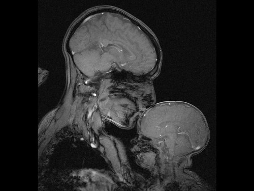
Neuroscientist captures an MRI of a mother and child
Professor Rebecca Saxe (MIT) has taken the first ever MR image of a mother and child.
“This picture is an MR image of a mother and a child that I made in my lab at MIT. You might see it as sweet and touching… an image of universal love. We can’t see clothes or hairstyles or even skin colour. From what we do see, the biology and the brains, this could be any mother and child or even father and child at any time and place in history; having an experience that any human can recognise.
Or you might see it as disturbing, a reminder that our human bodies are much too fragile as houses for ourselves. MRI’s are usually medical images and often bad news. Each white spot in that picture is a blood vessel that could clog, each tiny fold of those brains could harbour a tumour. The baby’s brain maybe looks particularly vulnerable pressed against the soft thin shell of its skull.
I see those things, universal emotions and frightening fragility but I also see one of the most amazing transformations in biology.”
Quotes have been taken from a TEDx talk given by Professor Saxe explaining the story behind the above picture.

Genes to Brains
Over the years scientists have carefully mapped the brain, figuring out which regions perform different functions. Techniques such as functional MRI can show exactly which parts are active when people are doing all kinds of other tasks. Detailed microscopy and brain-scanning studies have traced the intricate network of connections between nerve cells in the brain, revealing the inner wiring of this powerful biological computer. But until now, nobody has tried to link patterns of gene activity into this functional and structural information. For the first time, researchers have generated this map of the brain, with each colour highlighting a particular group of genes that seem to be linked to that region. There are many variations in human genes that can influence traits and conditions affecting the brain, such as intelligence or autism, and this is the first step towards figuring out exactly how these genetic variations might exert their effects.
Written by Kat Arney
Image from work by Qian Peng and colleagues
Department of Human Biology, J. Craig Venter Institute, and Multimodal Imaging Laboratory, Department of Radiology, University of California San Diego, La Jolla, CA, USA
Image originally published under a Creative Commons Licence (BY 4.0)
Published in PLOS Genetics, July 2016
You can also follow BPoD on Twitter and Facebook
Toxic Alzheimer’s Protein Spreads Through Brain Via Extracellular Space
A toxic Alzheimer’s protein can spread through the brain—jumping from one neuron to another—via the extracellular space that surrounds the brain’s neurons, suggests new research from Karen Duff, PhD, and colleagues at Columbia University Medical Center.

(Image caption: Orange indicates where tau protein has traveled from one neuron to another. Credit: Laboratory of Karen Duff, PhD)
The spread of the protein, called tau, may explain why only one area of the brain is affected in the early stages of Alzheimer’s but multiple areas are affected in later stages of the disease.
“By learning how tau spreads, we may be able to stop it from jumping from neuron to neuron,” says Dr. Duff. “This would prevent the disease from spreading to other regions of the brain, which is associated with more severe dementia.”
The idea the Alzheimer’s can spread through the brain first gained support a few years ago when Duff and other Columbia researchers discovered that tau spread from neuron to neuron through the brains of mice.
In the new study, lead scientist Jessica Wu, PhD, of the Taub Institute discovered how tau travels by tracking the movement of tau from one neuron to another. Tau, she found, can be released by neurons into extracellular space, where it can be picked up by other neurons. Because tau can travel long distances within the neuron before its release, it can seed other regions of the brain.
“This finding has important clinical implications,” explains Dr. Duff. “When tau is released into the extracellular space, it would be much easier to target the protein with therapeutic agents, such as antibodies, than if it had remained in the neuron.”
A second interesting feature of the study is the observation that the spread of tau accelerates when the neurons are more active. Two team members, Abid Hussaini, PhD, and Gustavo Rodriguez, PhD, showed that stimulating the activity of neurons accelerated the spread of tau through the brain of mice and led to more neurodegeneration.
Although more work is needed to examine whether those findings are relevant for people, “they suggest that clinical trials testing treatments that increase brain activity, such as deep brain stimulation, should be monitored carefully in people with neurodegenerative diseases,” Dr. Duff says.

A French man who lives a relatively normal, healthy life - despite missing 90 percent of his brain - is causing scientists to rethink what it is from a biological perspective that makes us conscious.
Despite decades of research, our understanding of consciousness - being aware of one’s existence - is still pretty thin. We know that it’s somehow based in the brain, but then how can someone lose the majority of their neurons and still be aware of themselves and their surroundings?
First described in The Lancet in 2007, the case of the man with the missing brain has been puzzling scientists for almost 10 years.
Read more…


Giant Artwork Reflects The Gorgeous Complexity of The Human Brain
The new work at The Franklin Institute may be the most complex and detailed artistic depiction of the brain ever.
Your brain has approximately 86 billion neurons joined together through some 100 trillion connections, giving rise to a complex biological machine capable of pulling off amazing feats. Yet it’s difficult to truly grasp the sophistication of this interconnected web of cells.
Now, a new work of art based on actual scientific data provides a glimpse into this complexity.
The 8-by-12-foot gold panel, depicting a sagittal slice of the human brain, blends hand drawing and multiple human brain datasets from several universities. The work was created by Greg Dunn, a neuroscientist-turned-artist, and Brian Edwards, a physicist at the University of Pennsylvania, and goes on display at The Franklin Institute in Philadelphia.
“The human brain is insanely complicated,” Dunn said. “Rather than being told that your brain has 80 billion neurons, you can see with your own eyes what the activity of 500,000 of them looks like, and that has a much greater capacity to make an emotional impact than does a factoid in a book someplace.”
To reflect the neural activity within the brain, Dunn and Edwards have developed a technique called micro-etching: They paint the neurons by making microscopic ridges on a reflective sheet in such a way that they catch and reflect light from certain angles. When the light source moves in relation to the gold panel, the image appears to be animated, as if waves of activity are sweeping through it.
First, the visual cortex at the back of the brain lights up, then light propagates to the rest of the brain, gleaming and dimming in various regions — just as neurons would signal inside a real brain when you look at a piece of art.
That’s the idea behind the name of Dunn and Edwards’ piece: “Self Reflected.” It’s basically an animated painting of your brain perceiving itself in an animated painting.
To make the artwork resemble a real brain as closely as possible, the artists used actual MRI scans and human brain maps, but the datasets were not detailed enough. “There were a lot of holes to fill in,” Dunn said. Several students working with the duo explored scientific literature to figure out what types of neurons are in a given brain region, what they look like and what they are connected to. Then the artists drew each neuron.
Dunn and Edwards then used data from DTI scans — a special type of imaging that maps bundles of white matter connecting different regions of the brain. This completed the picture, and the results were scanned into a computer. Using photolithography, the artists etched the image onto a panel covered with gold leaf.
“A lot of times in science and engineering, we take a complex object and distill it down to its bare essential components, and study that component really well” Edwards said. But when it comes to the brain, understanding one neuron is very different from understanding how billions of neurons work together and give rise to consciousness.
“Of course, we can’t explain consciousness through an art piece, but we can give a sense of the fact that it is more complicated than just a few neurons,” he added.
The artists hope their work will inspire people, even professional neuroscientists, “to take a moment and remember that our brains are absolutely insanely beautiful and they are buzzing with activity every instant of our lives,” Dunn said. “Everybody takes it for granted, but we have, at the very core of our being, the most complex machine in the entire universe.”
Image 1: A computer image of “Self Reflected,” an etching of a human brain created by artists Greg Dunn and Brian Edwards.
Image 2: A close-up of the cerebellum in the finished work.
Image 3: A close-up of the motor cortex in the finished work.
Image 4: This is what “Self Reflected” looks like when it’s illuminated with all white light.
Image 5: Pons and brainstem close up.
Image 6: Putkinje neurons - color encodes reflective position in microetching.
Image 7: Primary visual cortex in the calcarine fissure.
Image 8: Basal ganglia and connected circuitry.
Image 9: Parietal cortex.
Image 10: Cerebellum.
Credit for all Images: Greg Dunn - “Self Reflected”
Source: The Huffington Post (by Bahar Gholipour)


